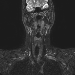
Head
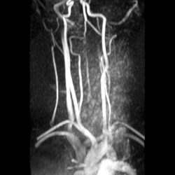
CT and MRI images of the brain, head vessels, and temporomandibular joints
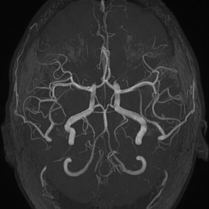
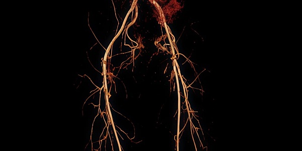
Arteries of the brain
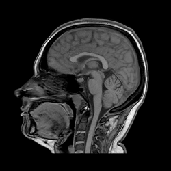
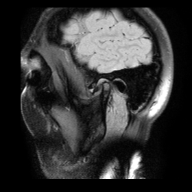
Brain
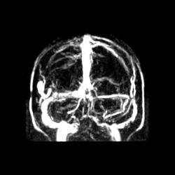
Head veins
Temporomandibular joints
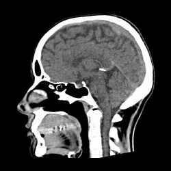
Brain
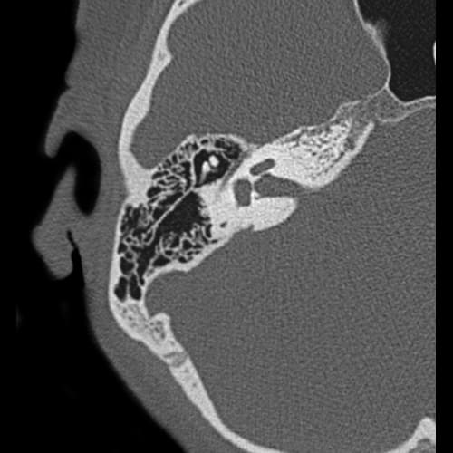
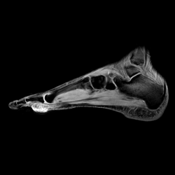
Temporal bones
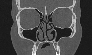
Paranasal sinuses
Neck
CT and MRI images of the neck soft tissues and arteries, and the brachial plexus
Vessels of the neck
Soft tissues of the neck
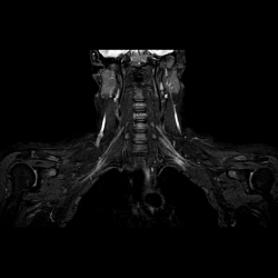
The brachial plexus
Thorax
CT and MRI images of the heart, lungs, and mediastinum, and the breasts
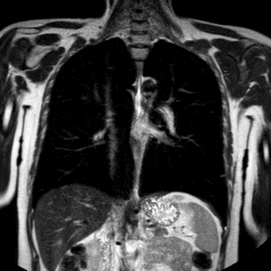
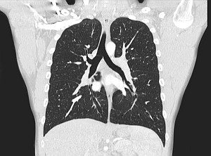
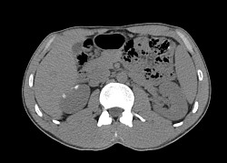
Lungs and Mediastinum
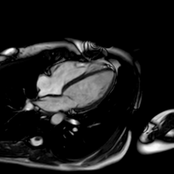
Heart
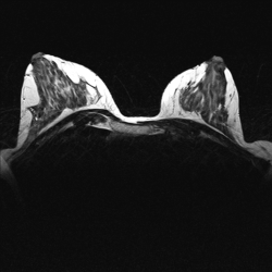
Mammary glands
Chest organs
Spine
CT and MRI images of the spine and the sacrococcygeal region, including the sacroiliac joint
Sacrococcygeal spine

Thoracic spine
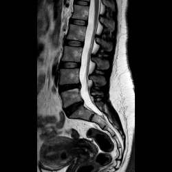
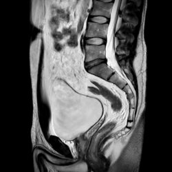
Lumbar spine
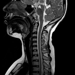
Cervical spine
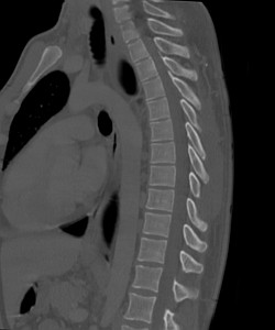
Thoracic spine
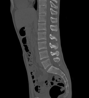
Lumbar spine
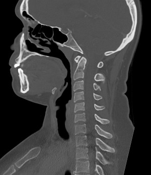
Cervical spine
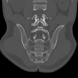
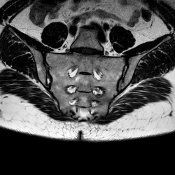
Sacrococcygeal region
Abdomen and pelvis
CT and MRI images of the abdominal organs and retroperitoneum, pelvic organs, and the male external genitalia
Abdominal cavity and retroperitoneal space
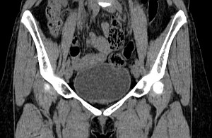
Male external genitalia
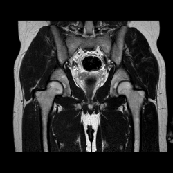
Male pelvis
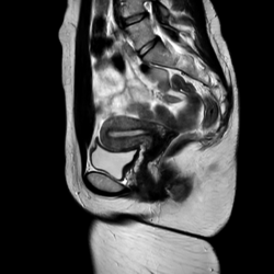
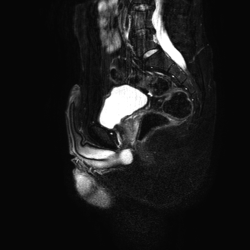
Female pelvis
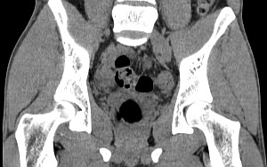
Male pelvis
Female pelvis
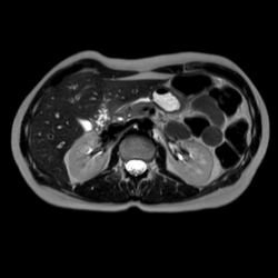
Abdomen
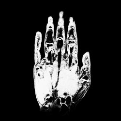
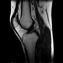
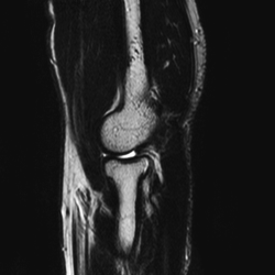
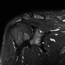
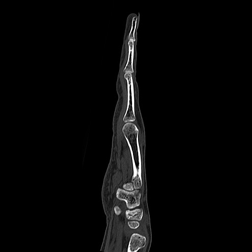
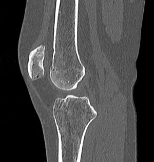
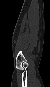
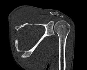
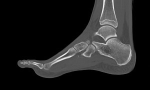
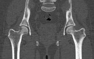
Upper and lower limbs
CT and MRI images of the joints of the upper and lower limbs
About the Project
The Radiological Anatomy Atlas is an interactive reference and educational tool for studying normal anatomy on CT and MRI images. Materials are organized by anatomical region, while manually labeled structures help users navigate slices faster, review anatomical landmarks, and work with images in a format close to real radiology practice.
How to Use the Atlas
Choose an anatomical region, open a CT or MRI section, and study the images slice by slice. Turn anatomical labels on and off, switch between label languages, and use the Atlas as a learning resource, a reference tool for reviewing anatomy, or visual material for teaching students and residents.